22 Jul | 2024
Cerebral blood flow decreases in asymptomatic stages of Alzheimer’s

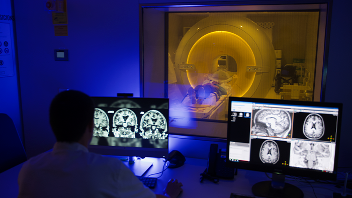
A multilateral collaboration led by the Barcelonaβeta Brain Research Center (BBRC), the research center of the Pasqual Maragall Foundation, has been able to measure, thanks to a new Magnetic Resonance Imaging (MRI) sequence, a decrease in the cerebral blood flow in the very first stages of Alzheimer’s disease, before the clinical symptoms appear. The project has involved experts in the development of novel MRI sequences, clinical professionals at the Hospital del Mar and collaborators providing state-of-the-art biomarkers of Alzheimer’s disease.
The team has used a new technique, Time-encoded Arterial Spin Labeling (teASL), to detect the very early changes in the cerebral blood flow of the study participants. The results of the study, published in the scientific journal Alzheimer’s & Dementia, show that people affected by the pathology of the disease also display lower blood flow in specific areas of the brain during its first stages.
Measuring the reduction
One of the first processes that are activated in the brain due to the presence of Alzheimer’s pathology (that is, an accumulation of amyloid beta and tau proteins), is a decrease in the cerebral blood flow. Blood supplies oxygen and glucose to the brain and, therefore, needs to be maintained within normal levels to ensure the brain’s health and proper functioning. Alterations in cerebral blood flow can precede or accompany various neurological conditions, including Alzheimer’s disease and, as such, accurately measuring it is vital for understanding these conditions. Arterial Spin Labeling (ASL) techniques allow to measure the cerebral blood flow using MR pulse sequences.
“Until now, the ASL techniques available allowed us to measure and compare cerebral blood flow in people with single delay time. This refers to the time it takes for the arterial blood to transit from the carotid arteries (where it is tagged) to the brain region of interest (known as arterial transit time)”, explains Dr. Michalis Kassinopoulos, postdoctoral researcher at the BBRC and one of the main authors of the study.
Thanks to a research collaboration with Philips, the BBRC has had access to a new ASL MRI sequence developed by Leiden University Medical Center, known as time-encoded ASL (teASL). This is a more sensitive and accurate tool reducing the intra-subject arterial transit time differences in the estimation of the cerebral blood flow. Researchers have used teASL to measure cerebral blood flow and investigate its association with amyloid and tau pathology, both of which are implicated in Alzheimer’s disease. Additionally, they have examined the relationship of decreases in cerebral blood flow with biomarkers in cerebrospinal fluid related to synaptic dysfunction and neurodegeneration, as well as cognitive performance. This way, the study has demonstrated, for the first time in asymptomatic individuals, that the levels of cerebral blood flow are associated with markers of tau pathophysiology, synaptic dysfunction and neurodegeneration.
Findings to define future prevention strategies
For this study, a total of 59 participants were separated into three groups: 24 healthy participants without cognitive impairment or amyloid protein accumulation in the brain (the “control” group); 18 healthy volunteers without cognitive impairment but with amyloid pathology present, and 17 patients from the Medical Research Unit at the Hospital del Mar in Barcelona, affected by the disease. Out of the healthy participants, around 30 belong to the Alfa study, promoted by “la Caixa” Foundation.
The study provides evidence that a reduced cerebral blood flow is not only present in persons in symptomatic Alzheimer’s stages, but also in cognitively unimpaired individuals harboring cerebral amyloid-beta pathology. “Reduced cerebral blood flow is an earlier event in the pathological cascade than previously thought, spanning preclinical stages”, asserts Dr. Juan Domingo Gispert, collaborator of the BBRC and corresponding author of the study. “These findings provide insight into the role of this early process in the disease, and can help shape future prevention strategies”, he concludes.
Reference
Falcon C, Montesinos P, Václavů L, Kassinopoulos M, et al. Time-encoded ASL reveals lower cerebral blood flow in the early AD continuum. Alzheimer's Dement. 2024; 1-15. https://doi.org/10.1002/alz.14059


